
How Does Pepstatin Inhibit Proteases? From Transition-State Mechanism to Purification Applications
Published on October 28, 2026
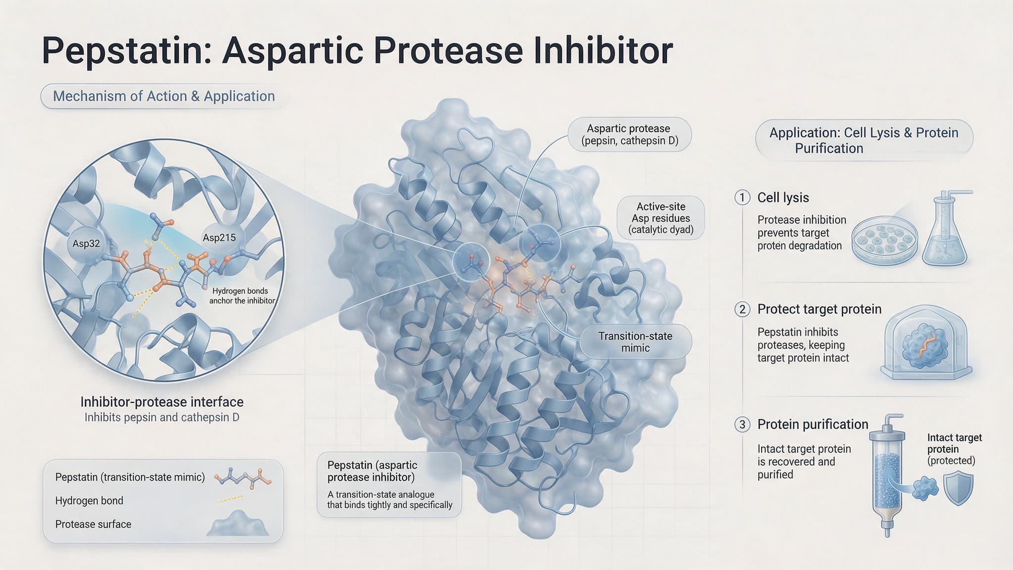
figure1. The overall scene of pepstatin binding to the aspartic protease active site
Category: Enzyme/Proteolysis | Application/Scenario (Primary A; Secondary G)
A target protein that keeps "disappearing" during purification is often not an expression problem but the work of endogenous proteases. Pepstatin, an aspartic protease inhibitor, mimics the transition state of peptide-bond hydrolysis, binds tightly to the catalytic aspartate dyad of the protease, and blocks the degradation of the target protein by pepsin and cathepsin D at picomolar concentrations. Understanding its inhibitory mechanism and proper use is a key step toward stable yields and lower batch-to-batch variability.
1. Pain-Point Approach: Why the Target Protein Keeps Disappearing
1.1 Endogenous proteases are the "invisible killer" of protein degradation
After cell disruption, endogenous proteases that were compartmentalized are released in large amounts and become the main cause of target-protein degradation. During cell lysis, tissue homogenization or purification, endogenous aspartic proteases (such as cathepsin D) act on the target protein together with serine and cysteine proteases, causing fainter bands, fragmentation and lower activity. Without inhibition, even a very high expression level may yield a disappointingly low final recovery.
The degradation usually shows up as smeared or ladder-like bands on an electrophoresis gel, a fainter target band, and lower activity or yield. If the target protein is protease-sensitive or the system is rich in cathepsins, even optimized expression may still yield only fragmented protein at harvest, affecting downstream purification and functional studies.
1.2 Breaking down the pain: technical, cost and compliance impact
Protein degradation simultaneously raises technical difficulty, amplifies cost and affects result compliance. Technically, degradation introduces heterogeneity, widening purification peaks, increasing impurities and reducing reproducibility; in cost terms, less usable protein forces repeat expression and larger batches, raising reagent and labor spend; for compliance, in analysis or filing scenarios requiring "intact and homogeneous" material, degradation fragments interfere with quality control and quantitation. Controlling degradation is therefore a foundational capability to build early in R&D.
1.3 Why adding inhibitors blindly is not always effective
An inhibitor is not "more is better"; the wrong choice or use can render it ineffective or even interfere downstream. Different proteases require matched inhibitors; the wrong category misses the main degradation source; an unsuitable concentration, solvent or addition timing may inactivate the inhibitor or affect later detection. The first step is to identify "what is degrading the protein," then inhibit it specifically.
2. Underlying Principle: How Pepstatin Locks Aspartic Proteases
2.1 The catalytic aspartate dyad of aspartic proteases
The active center of aspartic proteases (such as pepsin and cathepsin D) contains two aspartate residues that hydrolyze the peptide bond through general acid-base catalysis. The two aspartates act together, one as a base abstracting a proton from water and the other as an acid stabilizing the transition state, forming a tetrahedral intermediate before the peptide bond is cleaved. This highly conserved "aspartate dyad" is precisely the target for inhibitor design.
This catalytic mechanism makes aspartic proteases highly sensitive to spatial and electrostatic environment, which also explains why a molecule that closely mimics the transition state can achieve very strong inhibition. Pepstatin exploits this by binding the active site in a 1:1 stoichiometry, forming a high-affinity, near-irreversible complex.

figure2. The transition-state mimicry mechanism of pepstatin
2.2 Transition-state mimicry: the inhibitory mechanism of pepstatin
Pepstatin mimics the transition state of peptide-bond hydrolysis and forms a stable 1:1 complex with the catalytic aspartate dyad, thereby blocking catalysis. It is a hexapeptide in which the statine (Sta) residue carries a hydroxyl that closely reproduces the spatial and electronic features of the tetrahedral intermediate, binding tightly to the active site and preventing the enzyme from completing normal catalysis. This mechanism gives pepstatin high inhibitory potency against aspartic proteases.
Besides pepsin and cathepsin D, renin, chymosin and Aspergillus protease B are also aspartic proteases and can all be inhibited by pepstatin. It is therefore used not only to protect proteins but also as a tool to study aspartic-protease function, modulate autophagy and lysosomal pathways, or in experiments that require inhibiting this class of enzymes.
2.3 Why the statine (Sta) residue is key to inhibition
The statine (Sta) residue is the core of pepstatin's potent inhibition. It is not a common amino acid but a special residue bearing a hydroxyl that mimics the transition-state intermediate and forms a stable hydrogen-bond network with the two aspartates. This is why pepstatin forms a high-affinity 1:1 complex, with inhibitory potency reaching the picomolar range. In sequence, pepstatin is isovaleryl (Iva)-Val-Val-Sta-Ala-Sta.
2.4 Selectivity: why it acts only on aspartic proteases
Pepstatin's selectivity arises from the match between its transition-state mimicry and the catalytic mechanism of aspartic proteases. Because only aspartic proteases possess this aspartate dyad, pepstatin is highly specific to them and has little effect on serine or cysteine proteases. This makes it suitable as a "targeted" inhibitor of aspartic proteases, protecting target proteins prone to cathepsin-mediated degradation.
3. Evaluation and Selection: Metrics, Combinations and Boundaries
3.1 Key metrics for evaluating inhibitory potency
Evaluating an inhibitor mainly involves inhibitory potency (such as IC50), binding strength (dissociation constant) and whether a 1:1 complex forms. Pepstatin reaches nanomolar IC50 against aspartic proteases such as pepsin and binds in a 1:1 stoichiometry. In practice, the result should be judged together with the degradation level of the target system (band integrity, activity recovery) rather than by a single number.
For evaluation, use SDS-PAGE to check band integrity, activity assays for recovery, and if needed mass spectrometry or N-terminal sequencing to confirm whether the protein has been cleaved. Comparing before and after inhibition makes it clearer whether the inhibitor truly improves target-protein stability.
3.2 Division of labor among pepstatin and other protease inhibitors
Different inhibitors have distinct roles; pepstatin covers aspartic proteases, while serine, cysteine and metalloproteases require separate inhibitors. In a common cocktail, AEBSF/PMSF inhibit serine proteases, leupeptin covers serine and cysteine proteases, E-64 inhibits cysteine proteases, EDTA inhibits metalloproteases, and pepstatin A addresses aspartic proteases. Combining them as needed provides broad-spectrum protection.
Practical use must also balance compatibility with the downstream system. Solvents such as DMSO or ethanol may affect certain downstream assays or enzyme activity, so the final solvent concentration should be controlled. In addition, different inhibitors vary in their sensitivity to pH, temperature and metal ions; when preparing a cocktail, confirm the components are compatible and do not inactivate each other.
3.3 When a "cocktail" is necessary and when it can be simplified
The more complex the system and the more prone to degradation the target protein, the more a broad-spectrum cocktail is needed; when the system is clear and dominated by a single protease, it can be simplified. Mammalian cells and tissue homogenates usually have complex protease profiles and favor broad-spectrum combinations; in yeast, bacteria or later purification stages, once the degradation source is known, only the targeted inhibitor may be kept to reduce interference. The basis for the decision is the measured degradation pattern, not the assumption that "more is safer."
4. How to Benefit from MATWINGS MALL Products and Custom Services
Standard inhibitors/proteases cannot fit specific systems? MATWINGS MALL custom tool-enzyme development
When off-the-shelf inhibitors or general-purpose proteases cannot stably cover endogenous degradation in a particular system, researchers can rely on the custom tool-enzyme development service offered by MATWINGS MALL to develop toward target traits. Facing special hosts, complex substrates or special process conditions, MATWINGS MALL supports wild enzyme/protein mining, directed engineering and multi-objective performance optimization, and can develop adapted variants as needed, supported by dry-wet integrated validation and sample evaluation to help lower trial-and-error cost and let candidates be assessed verifiably before scale-up.
5. Practical Recommendations and Pitfalls

figure3. A protease inhibitor cocktail containing pepstatin across cell lysis, protein extraction
5.1 Preparation, storage and solubility notes
Pepstatin is nearly insoluble in water and is usually first dissolved in methanol, ethanol or DMSO containing acetic acid before use. Follow the instructions to dissolve it in a suitable solvent to avoid precipitation when added to an aqueous phase; store under the recommended conditions, such as low temperature and protection from light, to prevent degradation or inactivation.
If the system also has metalloproteases or metal-ion interference must be avoided, an EDTA-free formulation can be used, or a judicious choice made on chelators. The final concentration of the introduced DMSO or ethanol should not be too high, to avoid affecting protein conformation or downstream activity assays.
5.2 Addition timing and concentration
The inhibitor should be added before or at the moment of protein release, and at a sufficient concentration. It is advisable to pre-mix the inhibitor in the lysis buffer so the protein is protected the instant cells break; use the recommended concentration range, since too low a level fails to inhibit fully and too high a level may interfere with downstream assays. The suitable concentration should be determined by a gradient measured in the actual system.
5.3 Pitfall checklist and troubleshooting
If degradation persists despite adding the inhibitor, troubleshoot in the order "solvent - inactivation - concentration - degradation source." First confirm whether the inhibitor was inactivated by improper solvent or storage; then check whether the concentration is sufficient and covers the main protease types; meanwhile run paired with/without inhibitor controls to locate the true degradation cause, and verify with electrophoresis and activity assays if needed.
6. FAQ
6.1 What type of protease inhibitor is pepstatin?
Pepstatin is an aspartic protease inhibitor that selectively inhibits aspartic proteases such as pepsin, cathepsin D/E and renin.
6.2 Why can pepstatin inhibit proteases?
It mimics the transition state of peptide-bond hydrolysis and forms a stable 1:1 complex with the catalytic aspartate dyad, thereby blocking catalysis.
6.3 Can pepstatin inhibit all proteases?
No, it mainly targets aspartic proteases and has little effect on serine or cysteine proteases, so it usually needs to be combined with other inhibitors.
6.4 How is pepstatin used in protein purification?
During cell lysis, tissue homogenization and purification, adding a protease inhibitor combination containing pepstatin blocks endogenous degradation and protects target-protein integrity.
6.5 Pepstatin is insoluble in water; how should it be prepared?
First dissolve it in methanol, ethanol or DMSO containing acetic acid, then add it to the system to avoid precipitation when added directly to an aqueous phase.
6.6 What if degradation still occurs after adding pepstatin?
Troubleshoot in the order "solvent - inactivation - concentration - degradation source," and run paired with/without inhibitor controls to identify the true cause.
7. Summary
The value of pepstatin lies in precisely locking aspartic proteases through transition-state mimicry, providing a layer of protection against endogenous degradation during protein extraction and purification. It is not universal, targeting only aspartic proteases and needing combination with other inhibitors for broad-spectrum protection; its potency stems from the stable binding between the statine (Sta) residue and the catalytic aspartate dyad. In practice, first locate the degradation cause, then choose the inhibitor combination for the system, and pay attention to preparation, concentration and addition timing. When standard solutions cannot cover a specific system, researchers can turn to the custom tool-enzyme development service from MATWINGS MALL to develop and validate toward target traits. For related products and services, see the MATWINGS MALL product zone and further industry insights.