Light-Chain-Binding Ligand: An Affinity Purification Guide
Published on September 3, 2026
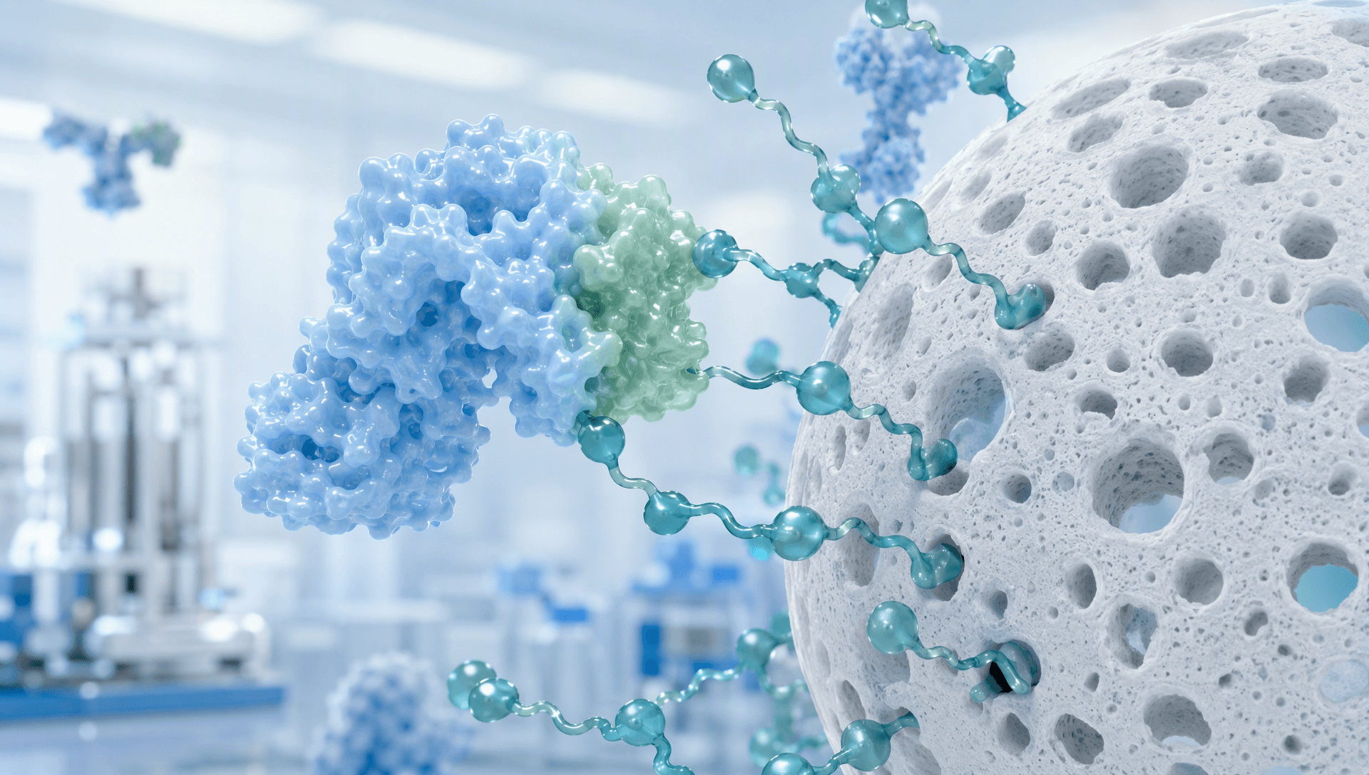
Figure 1 | A Light-Chain-Binding Ligand used for antibody-fragment affinity purification. The schematic does not represent the performance of a specific resin.
The main value of a Light-Chain-Binding Ligand is that it moves affinity recognition away from the Fc region and toward features associated with the light chain. Protein A and Protein G may lose their usual capture role when an intact Fc region is absent. A ligand that recognizes a light-chain structure can therefore offer an alternative for Fab or selected scFv molecules. Yet light-chain recognition is not equivalent to universal fragment compatibility: subgroup, local conformation, epitope exposure, and feed environment all shape process performance.
What does a Light-Chain-Binding Ligand recognize?
An antibody light chain contains variable and constant regions and pairs with a heavy chain to form the antigen-binding structure. Different ligands may recognize different surfaces. Some depend on a conformational site in the variable region, others target a narrower sequence or structural feature, and engineered proteins or peptides may be selected for a defined Fab family.
Protein L is a familiar example. It interacts with structures associated with certain kappa light chains and does not require an intact Fc region. This makes it relevant to some Fab and light-chain-containing scFv formats. Its mechanism differs from Protein A or Protein G, which are primarily used through Fc recognition.
The boundary matters. Species, V-kappa family, framework variation, and fragment conformation can change binding. Lambda-containing molecules should not automatically be treated as compatible with a kappa-directed ligand. VHH and other single-domain formats do not contain a conventional light chain, so the phrase “antibody fragment” alone is not enough to predict binding. A Light-Chain-Binding Ligand is therefore a mechanism class, not a universal tag.
Why one ligand behaves differently across molecules
Affinity chromatography is governed by the combined system of ligand, target, matrix, and buffer.
First, the binding site must remain accessible in the native construct. Two kappa-containing Fabs may differ in framework geometry, heavy-light pairing, or neighboring domains. Fusion partners and bispecific architectures can also create steric shielding.
Second, immobilization changes effective interaction. Low ligand density can limit capacity, whereas excessive density may increase crowding or multivalent binding and make elution more difficult. Pore architecture, particle size, and spacer length influence how efficiently a fragment reaches the ligand.
Third, the feed determines practical selectivity. pH, conductivity, host-cell proteins, surfactants, and other additives may alter target binding or nonspecific adsorption. Performance in a clean buffer cannot substitute for behavior in cell-culture harvest, lysate, or digestion material.
For that reason, ligand evaluation should include breakthrough loss, recovery, aggregation, retained activity, peak shape, and impurity clearance—not binding alone.

Figure 2 | Recognition, impurity flow-through, and controlled elution for an antibody light chain and an immobilized affinity ligand.
From Protein L to engineered affinity ligands
Native or recombinant Protein L offers a defined mechanism and direct capture for compatible kappa-containing Fab or scFv molecules. It is useful for rapid feasibility testing, but its coverage is limited and its elution conditions must be compatible with fragment stability.
Engineered protein ligands may be optimized for affinity, stability, alkaline cleaning tolerance, coupling efficiency, or milder release. Stronger binding is not automatically better. Excessive affinity can reduce recovery or require harsher elution. A process-ready ligand must bind reliably under loading conditions, release product under acceptable conditions, and retain performance during cleaning and cycling.
Peptide or small-molecule ligands provide additional design space and can be selected for one Fab or a related group. Platform claims require evidence across representative molecules; success with one construct does not establish broad compatibility.
Engineered purification tags are another practical route for recombinant fragments. They can accelerate early development, but tag retention, proteolytic removal, residual tag, and residual enzyme must be considered in the complete process. Tag capture is not identical to native light-chain recognition, although it can serve as an alternative when no suitable natural epitope is available.
How to screen a Light-Chain-Binding Ligand
Begin by confirming light-chain type, sequence context, molecular format, and epitope accessibility. Use a small-scale binding experiment with the actual target. Species or the broad label “Fab” is not sufficient to establish compatibility.
Next, test representative feed. Culture harvest, cell lysate, and antibody digest contain different impurities. A high binding signal with purified standard protein does not demonstrate selectivity in process material. Analyze load, flow-through, wash, and eluate to establish a mass balance.
Elution belongs in the initial screen. Low pH, salt, competitive additives, or other release conditions may affect recovery and stability. For aggregation-prone fragments, minimize exposure to unfavorable conditions and neutralize or exchange buffer promptly.
Scalability must also be evaluated. Dynamic binding capacity, residence time, pressure-flow behavior, cleaning, ligand leakage, and cycling decay determine whether a laboratory result can become a reproducible operation. When one affinity step does not meet quality goals, ion exchange, hydrophobic interaction, or size exclusion can provide orthogonal polishing.
The decision sequence should be explicit: first meet recovery, activity, and critical impurity limits; then compare capacity, buffer use, and medium cost. This prevents a high single-step purity value from hiding poor functional recovery or process robustness.
Supporting ligand-screening preparation with MatwingsVenus™(晓鹜™)

Figure 3 | Development workflow from molecular information and binding hypotheses to resin screening and process verification.
A concrete task chain starts with the fragment name, sequence, light-chain type, expression system, and priority impurities. The deep-research capability of MatwingsVenus™(晓鹜™)can return sourced summaries of mechanisms and public studies, separating established evidence from open questions. The protein-database query capability of MatwingsVenus™(晓鹜™)can organize sequence, domain, and known annotation data into a molecular information card. If measured properties are unavailable, the protein-property prediction capability of MatwingsVenus™(晓鹜™)may provide explicitly labeled hypotheses about pI or stability for designing pH, conductivity, and elution screens.
The next step is not automatic selection of a commercial resin. The outputs should become a candidate-ligand list, a small-column study, and a defined analytical panel. Binding, recovery, impurity clearance, cycling stability, and scale-up behavior still require experimental confirmation; digital analysis does not replace process validation or product release.
Selection principle
A Light-Chain-Binding Ligand can provide a valuable affinity route for antibody fragments without an intact Fc region, but its value comes from molecular fit rather than universality. Developers should confirm light-chain compatibility, selectivity in representative feed, elution impact on activity, matrix transport and cycling behavior, and the need for orthogonal polishing. Evaluating molecular recognition, chromatographic conditions, and scale-up constraints together is the route from an experimental ligand to a stable purification step.